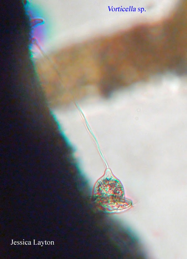
This week happens to be the final week of observations on my microquarium project, and sadly it did not end with a bang. The activity in my microquarium was about the same as last week, though arguably I saw more organisms floating around. This week I observed only a select few of the organisms found in my previous observations, such as Rotifers, Eulopes and a critter I had yet to identify, the Coleps sp. This organism in paticular has flourished despite the lack of population density of other species. For example I saw a massive group of over 10 individuals swimming around in circles. They also appeared in many other areas of the aquarium. Durring this exploration for life in the microaquarium I also noticed a few rotifer eggs, one of which was beggining to hatch. Regretibly I was not able to get a photograph of it, but I saw a few of these hatchlings throughout the aquarium as if the population was attempting to bounce back from last weeks sudden decline. Along side these stragglers, I saw a huge population of diatoms, however they too were struggling and had already lost their coloration in the somewhat unfavoring environment. Other than the diatoms, I did not find any new species in my microaquarium this week.
Here are the last few organisms I had left to identify:
The dominate species within the slowly decaying microaquarium. Go Little Coleps!
(Patterson 163)
This unusual organism was observed in the week 2 observations. Sadly I only observed about 3 individuals that week and never saw them in further observations.
(Pennak 218)
This organism was observed in weeks 1 and 2, this photograph in particular from the week 1 observations. I witnessed quite a few individuals, numbering over 20, between weeks 1 and 2. There was a tremendous increase in the population in week 2 when the food pellet was added, however in week 3 all organims dissapeared and I have not seen them since.
(Patterson 119)
As a final note, this project was quite interesting in that I really enjoyed seeing all of these microscopic organism that I would have never imagined previously. I observed quite a few organims and to process of discovering and identifying them was quite rewarding. I'm actally kind of sad its over.
Monday, November 17, 2014
Thursday, November 6, 2014
Week 3 Observations!
This week, nothing was added to the microaquariums. To my surprise however, my micro aquarium's activity decreased tremendously. All of the almost hundreds of rotifers and single celled organisms vanished, and only the remnants of plant material and multiple root structures filled the vacant void were bustling life had once filled. Despite the clear decrease in activity and organism population I was able to observe a few select organisms. I did come across one lone rotifer, swimming about in the open, a couple Litionotus very close to the soil bed, and a lone stentor unattached to any surface. In the absence of life however, I noticed a slight increase in the population of euplotes! I saw at least 10 different individuals, most hanging about near the soil bed. And finally, in the abscence of other critters, I discovered a new species inside my microaquarium that I had not previously seen as well as one other organism that I had seen on my first observation but had yet to classify. They are as follows:
(Patterson 54)
This little critter is a single celled organism with two flagella located at both ends of the the cell. This is the organism that I observed on week one, but hadn't seen in week two. The reason I waited to classify is because of my poor picture quality on week one, and as you can see, I acquired a pretty good picture this week :D
(Pennak 164-165)
This is the critter that I discovered this week, and I was actually pretty excited about! This one is by far, in my opinion, one of the most interesting critters I have found thus far. I saw a total of two individuals during my observations this week, and both were located among the vegetation or free floating/ swimming among the empty areas.
(Patterson 103)
These are three amoebae I found last week that I hadn't had the time to classify and post. I thought it very interesting that I found three in one area. These critters I have found sliding about in my microaquarium every week, week two being the peak of their population. I only came across one individual this week.
Though this week my aquarium had little life, I have hopes that next week the population may increase at least a little. My hope comes from the increase in plant life within the microaquarium. I found multiple strains of algae growing this week that may attract various individuals and provide food for the populations to grow. Next week I will be finishing my classifications of organisms that I have found over the course of these three weeks, as well a few organisms that I may find next week, and posting my final notes about the micro aquarium. I can't wait to see what I find next week!
Sunday, November 2, 2014
Week 2 Observations!
This past week I went in once again to investigate my micro aquarium and there was a lot to see! On October 24, Dr. McFarland added a single pellet of "Atison's Betta Food" made by Ocean Nutrition, Aqua Pet Americas, 3528
West 500 South, Salt Lake City, UT 84104. Ingredients: Fish meal, wheat
flower, soy meal, krill meal, minerals, vitamins and preservatives.
Analysis: Crude Protein 36%; Crude fat 4.5%; Crude Fiber 3.5%; Moisture
8% and Ash 15% (McFarland 2014). As a result the number of microorganims appeared to skyrocket. The litionotus sp. that I posted about last week was seen (not the same exact organism of course) quite a bit during my observations. I came across four or five that were mature and displayed the long neck like appendage. Also while looking about, I saw a wealth of very small unicellular organisms wiggling about in the background of the largest magnification almost everywhere! Upon investigating the food pellet itself I saw a huge amount of life, many organisms that I have yet to identify, but will definitely get into next weeks blog. The pellet iself attracted a multitude of organisms including these three organisms:
(Pennack 183)
(Patterson 124)
(Patterson 107)
Starting with the Echlanis Rotifer Sp. I have seen these critters in the previous observation and refered to them as reminding me of horseshoe crabs, of which they still do. Anyway, the sheer number of these little guys is astonishing and I would not be surprised if I have seen over 50 different individuals. They appear to stay close to vegetation and love to crawl all over it, joined by many of their fellow rotifers. Also they are quite quick, making it very difficult to capture a good picture of them free floating. They are most definitively the most prominent species I have seen in my microaquarium so far, other than the very small single cell organisms I mentioned earlier. Though it may not be noteworthy, they have also become my favorite critters in the micro aquarium too.
Next is the Eulotes sp. of which is one of the rarer micro organisms I have come across in my micro aquarium. I have come across only two thus far, one of which was found very close to vegetation and the one, that is in the picture above, that was free floating. This critter does not appear to be very fast and I was unable to capture one feeding on anything in particular.
The final picture is of a Stentor sp. I saw quite a few of these fascinating micro organisms in my microaquarium. I primarily saw them attached to vegetation and letting the upper portion of their body, as seen in the figure above, float with the flow of the water. I also found a single free floating Stentor which exhibited root like structures around the base that would be normally seen attached to some sort of vegetation. The blurred area around the 'head' so to speak are hundreds of moving flagella either sifting the water around it or keeping it in place. From the various individuals I saw, of which numbered over 10, they appear to be consuming smaller single celled organisms around them or floating particles of plant matter. Also it is note worthy to say that I saw an increase in the numbers of stentor around the food pellet, their numbers greatest in the higher micro organism traffic areas.
There were many other organisms that I captured on camera that I cannot wait to classify and share! Overall though, the total number of organisms increased two fold. I saw an abundance of activity almost everywhere, especially around the agitation and the food pellet. Its exciting to see my micro aquarium thriving and I cannot wait to see what I find next week!
Bibliography
Bibliography
McFarland, Kenneth [Internet] Botany 111 Fall 2014. [cited October21-November 13, 2014]. Available from http://botany1112014.blogspot.com/
Patterson, D.J. 1992. Free-Living Freshwater Protozoa. Manson Publishing Ltd. 54, 103, 107, 133, 124. 163pg.
Pennak, Robert W. 1989. Fresh-Water Invertebrates of the United States; Protozoa to Mollusca. 3rd ed. John Wiley & sons, Inc. 164-165, 183, 218pg.
Sunday, October 26, 2014
Observations of Week 1!
This week I observed my little micro aquarium under a different type of microscope that had the ability to magnify at a higher rate and take photographs. After a week of resting, the organisms within my microaquarium have begun to flourish and thrive in their new environment. Upon observing I can across many more single cell organisms than I had initially, and even began to see quite a few more complex single cell organisms. I saw quite a crustacean like micro organisms (they were not crustaceans but they reminded me of horseshoe crabs). I also saw quite a few simple rounded organisms and even an amoeba. Though I took many pictures, the one I have chose to focus on this week is of the Litonotus Sp. which I classified from the book Free-Living Freshwater Protozoa. Here is what he looks like:
Based on what the book says, this is a juvenile for of Litionous Sp. This is recognized by the lack of a "ingestion region is extended giving it the appearance of a long neck" (Patterson 133). These little critters are typically found in a water column, substrate or detritus, and their mouths tend to be very hard to see (Patterson 133). This was one of many creatures I caught on camera and I can't wait to begin classifying them and sharing them next week!
Bibliography:
Patterson, D.J. 1992. Free-Living Freshwater Protozoa. Manson Publishing Ltd. 133pg.
Tuesday, October 21, 2014
Building of the Micro Aquarium
Last week we began our micro aquarium term project, and during lab we built our very own micro aquariums using samples collected by Dr. McFarland. To build the aquarium itself, we did the following;
gathered the materials;
dat blue stick putty, a microquarium, a microaquarium lid, a micro aquarium base, and colored stickers to label our aquariums.
Then put them together by connecting the micro aquarium base to the microaquarium, then placed two beads of dat blue stick to the lid and placed that on top to create our very own micro aquarium!
Upon building these aquariums we made sure to include a viable soil sample, a few aquatic plants and water samples from various layers of the sample we chose. I chose to use sample 6. Once built I examined my aquarium under the microscope and didn't see very much at all. I spotted a few unknown single cell organisms swimming about and a very interesting one that spun around in circles sifting through the surrounding water. Once I removed the aquarium I also noticed a larger organism swimming around that I had not caught under the microscope. This organism was visible to the naked eye and a very light brown color. Dr. McFarland mentioned that it might be a cyclops. I hope I catch it under the microscope next time so I can see what this little critter looks like!
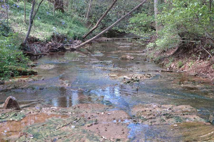
Also sample 6 was gathered at Dean's Woods Spring Creek. Here is a little more information on the area:

6. Spring Creek off Woodson Dr runing throught Dean's Woods Road frontage., Knox Co. Tennessee. Partial shade exposure. Tennessee River water Shed N35 55.274 W083 56.888 848 ft 10/12/2014
gathered the materials;
dat blue stick putty, a microquarium, a microaquarium lid, a micro aquarium base, and colored stickers to label our aquariums.
Then put them together by connecting the micro aquarium base to the microaquarium, then placed two beads of dat blue stick to the lid and placed that on top to create our very own micro aquarium!
Upon building these aquariums we made sure to include a viable soil sample, a few aquatic plants and water samples from various layers of the sample we chose. I chose to use sample 6. Once built I examined my aquarium under the microscope and didn't see very much at all. I spotted a few unknown single cell organisms swimming about and a very interesting one that spun around in circles sifting through the surrounding water. Once I removed the aquarium I also noticed a larger organism swimming around that I had not caught under the microscope. This organism was visible to the naked eye and a very light brown color. Dr. McFarland mentioned that it might be a cyclops. I hope I catch it under the microscope next time so I can see what this little critter looks like!
Also sample 6 was gathered at Dean's Woods Spring Creek. Here is a little more information on the area:

6. Spring Creek off Woodson Dr runing throught Dean's Woods Road frontage., Knox Co. Tennessee. Partial shade exposure. Tennessee River water Shed N35 55.274 W083 56.888 848 ft 10/12/2014
Subscribe to:
Comments (Atom)


